Laser Vision Treatment
Millions of people have chosen iLASIK™.
Jump to:
A Procedure as Unique as You
Because your eyes are as unique as your fingerprints, a one-size-fits-all approach to laser vision correction is not recommended.
After you've consulted with your ophthalmologist about iLASIK™, an extensive analysis – or a distinctive mapping – of your eyes is performed using the iDESIGN™ Refractive Studio.
This distinctive "map" of your eyes is produced, creating a complete analysis of the imperfections of your eye.
A Difference You Can Truly See
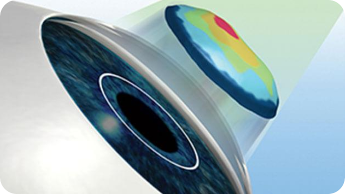
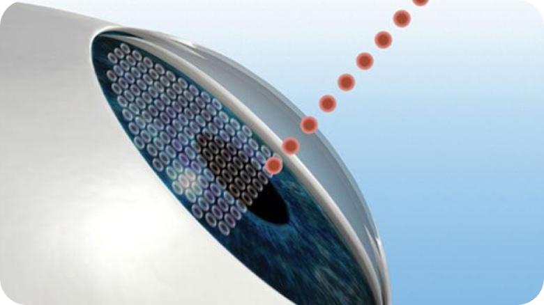
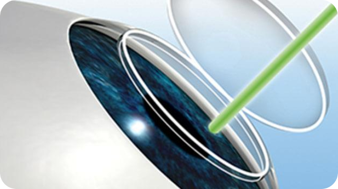
iDESIGN™ Refractive Studio technology takes over 1,200 measurements of your eye and maps each data point to create a custom iLASIK™ procedure plan designed just for you all in only three seconds.
iDESIGN™ Refractive Studio mapping offers 25 times more precision than conventional measurement,* resulting in 20/16† or better vision for the majority of myopia patients.**
*Based on mathematical calculation: wavefront aberrometer = 0.01 D; manifest refraction = 0.25 D. Manifest 0.25 ÷ wavefront aberrometer 0.01 = 25. 25X more precise.
**Based on FDA manufacturer excimer platform results, head to head studies, non-comparative studies, and podium abstracts & posters, iDESIGN™ has been shown to deliver 20/16 or better visual outcomes in majority of myopia patients. Data available upon request.
†FDA Study-P930016/S044
Preparation & Treatment
Mapping the Inner and Outer Eye
iDESIGN™ Refractive Studio maps both the inside and the outside of your eye.
The wavefront guided technology measures how light travels inside your eye and creates what you see, detailing the imperfections of your vision. It maps the entire optical pathway inside the eye.
In addition, the outer surface of your cornea is captured by scanning the outside of your eye, measuring every tiny variation in curvature and elevation.
Using this customized eye map of both the inner and outer eye, your ophthalmologist identifies optical imperfections that are affecting the quality of your vision, to be corrected during the iLASIK™ procedure.
Your Eye Map
As part of your customized eye map, your ophthalmologist also evaluates:
- Prescription stability
- Corneal health, shape, and thickness
- Tear layer
- Pupil size
- Existing conditions and their severity
Procedure
Clear Vision, in Less than 5 Minutes
Using your custom procedure plan created by the iDESIGN™ Refractive Studio system, the iLASIK™ procedure is able to correct your vision in less than 5 minutes, if you're getting both eyes done. In total, you can expect to be at the surgeon’s office for about an hour.
You cannot drive yourself home post-op, because your vision will be blurry. Depending on whether you were given a sedative, you may be a bit drowsy.
When you get home, the best way to begin healing is to nap or rest, and use recommended drops to keep your eyes moist, healthy and comfortable. It's common for full healing to take three months or longer. Ask your LASIK surgeon about recovery time.
No one in the world will receive the same procedure. Once the procedure is over, you'll notice the difference immediately.
During the prep stage, the iDESIGN™ Refractive Studio system uses an ultra-fast laser to prepare your cornea for treatment, which is an important step in the overall procedure.
How the iLASIK™ Treatment Process Works
Amazingly, the actual iLASIK™ procedure only takes about 20 minutes, and you’ll be awake the whole time. When you arrive for your appointment, your surgeon may provide medication to help you relax, as well as numbing eye drops to keep you comfortable.
Don't worry about blinking during the procedure. A small device will be used to hold your eyelids open. While the laser is active, an automated "eye tracker" monitors the position of your eye and compensates for any small, involuntary eye movements to deliver targeted treatment.
Preparing the eye
The procedure uses an ultra-fast laser to create a thin, customized flap that is gently folded back to prepare your cornea for treatment.
Delivering your personalized treatment
An ultra-precise laser is used to gently reshape your cornea to the desired curvature, based on the digital information from the iDESIGN™ Refractive Studio system's customized eye map.
Innovative Technology
Based on the digital information from your personalized eye map, an ultra-precise laser is used to gently reshape your cornea to the desired curvature. While the laser is active, an automated "eye tracker" monitors the position of your eye and compensates for any small, involuntary eye movements to deliver targeted treatment.
After one hour of having the iLASIK™ procedure, you'll begin to see more clearly.
How You May See After
Select a number based on your current visual acuity, and compare the potential post procedure results.
Within minutes after the iLASIK™ procedure, Brandi was already seeing more clearly, and now enjoys freedom from wearing contacts.*
*Individual results may vary
Post-Treatment
The Proof is in the Post-Op
At 6 months post-procedure, 323 nearsighted clinical study patients reported significant improvements in all measures of vision quality*:
Clarity of vision
Satisfaction with correction
Near vision
Far vision
Activity limitations
Difficulty driving at night
Benefits You Can See
Imagine going about your day without the hassle of glasses or contact lenses.** Ninety-four percent of patients see 20/20 or better† six months after laser vision correction. Before you know it, you'll be back to your normal daily routine.
*FDA P930016 supplements 044, 045 & 048.
**Some activities may require glasses.
†Based on clinical studies.
What to Expect Post-Op
It’s possible to experience issues such as dry eyes or glare after any type of corneal refractive procedure. iLASIK™ cannot stop your eyes from aging or from potentially needing reading glasses. Some activities may require glasses.
Now that you've had your vision improved through iLASIK™, you'll want to know how to maintain your eye health and safeguard your sight.
You cannot drive yourself home post op, because your vision will be blurry. Depending on whether you were given a sedative, you may be a bit drowsy.
When you get home, the best way to begin healing is to nap or rest, and use recommended drops to keep your eyes moist, healthy and comfortable.
It's normal to feel some moderate pain and discomfort for up to three days.
Some people report feeling something like an eyelash in the eye. As the cornea heals, you may experience blurriness, itchy, dry or watery eyes, and light sensitivity. These symptoms should subside over time.
Protecting Your Eyes
Protective shields will be placed over your eyes to help you avoid rubbing, touching, or bumping your eyes. In the days following your procedure, always wear sunglasses to shield your eyes from the sun and reduce glare. It's vital to keep your eyes clean and hydrated to optimize your recovery rate, but keep in mind it's common for full healing to take three months or longer.
Looking Forward
Generally, your first follow-up visit will be approximately 24 hours post-procedure. Even if your vision seems clear, have someone drive you to this visit. Your doctor will let you know when it's safe for you to drive again, and when you can resume other activities like most exercise. Remember that routine comprehensive eye exams are still absolutely needed to safeguard the health of your eyes.
If you experience significant discomfort after your iLASIK™ procedure, particularly if it worsens over time, call your ophthalmologist for further instructions.
Dry Eye Treatment Alleviates Symptoms
It’s been shown that Dry Eye caused by Meibomian Gland Dysfunction (MGD) can cause unsatisfactory vision and affect the overall results of cataract surgery.1 Dry eye symptoms caused by MGD tend to worsen with surgical procedures if they are not treated.
The good news is that the TearScience™ LipiFlow™ treatment can help optimize the ocular surface by stabilizing the tear film prior to surgery.2 In fact, in a recent study, patients treated with TearScience™ LipiFlow™ had a twofold reduction in dry eye symptoms 3 months after surgery.2
References:
1. Starr CE, Gupta PK, Farid M, et al. An algorithm for the preoperative diagnosis and treatment of ocular surface disorders. J Cataract Refract Surg. 2019 May;45(5): 669-684.
2. Data on file, 2018 (NCT01808560). Pilot study for treatment of meibomian gland dysfunction (MGD) prior to cataract surgery. Accessed from https://clinicaltrials.gov/show/NCT1808560.
PP2023OTH5324